DENTAID EXPERTISE
News for dentistry professionals
SURGICAL TREATMENT OF ORO-SINUSAL COMMUNICATIONS. A CLINICAL CASE
26 Apr 2019

From a clinical case, we present the benefits of a palatal rotational flap (PRF) as treatment for the closure of an oro-sinusal communication.
Ángela Martín Gómez, Andreína García de Frenza, Tatiana Ortiz Alves, Luis Oliveros López, Antonio Batista Cruzado, Daniel Torres Lagares. Master of Oral Surgery, School of Dentistry, Universidad de Sevilla.
INTRODUCTION
Oro-sinusal, or oro-antral, communication is a pathological condition where there is a solution of continuity between the oral cavity and the maxillary sinus(1). These structures can often be affected by infectious, cystic and/or mechanical traumatic processes, such as conventional extraction, in which upon extracting the roots, the two anatomical regions are left open to one another.
When oro-sinusal communication exceeds 4 mm, surgical treatment of the condition must be considered in order to minimise the risk of invasion of the maxillary sinus by oral bacteria, which may cause mild pain, swelling or even acute sinusitis, with such symptoms eventually becoming chronic(1).
To this end, there is a broad range of therapeutic options, such as biocompatible material grafts to impede the passing of saliva and food into the maxillary sinus(2-3), replacement flaps to allow first-intention healing of the tissue surrounding the communication, or connective tissue grafts from different areas of the oral cavity(4), amongst others.
AIM
The aim of this paper is to share the therapeutic option of the closure of oro-sinusal communications with the presentation of a clinical case resolved by means of a palatal rotational flap (PRF) that favours and enables the primary closure of the communication.
CASE DESCRIPTION
A 28-year-old male patient is referred to the Master in Oral Surgery at the School of Dentistry of the Universidad de Sevilla presenting root fragments remaining in the second upper left premolar. There is no medical history of interest. The patient reports episodes of mild pain and intermittent infection lasting several months.
Cone beam computed tomography (CBCT) reveals the presence of an apical cyst associated with the root fragments, closely related to the anterior/inferior wall of the left maxillary sinus with fenestration of the vestibular table at the apex (Figures 1, 2 and 3).
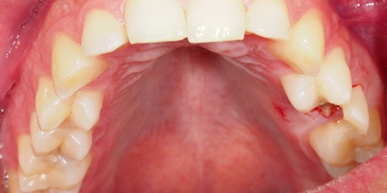
Upon inspection, root fragments with retained vestibular wall of the tooth are observed, with no other suspect remains (figure 4).
SURGICAL TECHNIQUE
With prior asepsis of the operating field and under local anaesthesia by infiltration of lidocaine 2% with epinephrine 1: 100,000 IU (Figure 5), extraction of the root remains (Figures 6 and 7), and removal of the lesion (Figure 8) are performed. Bearing in mind that there is a close relationship with the floor of the maxillary sinus, meticulous curettage of the area is carried out. Despite this, oro-sinusal communication arises, confirmed by a Valsalva manoeuvre, and is treated by PRF.
Prior to PRF, a vestibular flap is raised to show the bone defect in the vestibular table, favouring proper removal of any tissue that might interfere with subsequent healing of the area (Figure 9).
PRF surgery helps close the region properly. It begins with an incision in the palatal mucosa to lift a partial-thickness flap and rotate it on its pedicle in order to occlude the defect in the alveolar crest where it will be sutured (Figure 10). The donor site is left to bleed in order to allow second-intention healing (figures 11 and 12).
Afterwards, the patient is instructed in measures of postoperative care, and prescribed antibiotics, analgesics and anti-inflammatory medications for seven days(5). Checks are performed at 2, 8, and 12 weeks to verify proper healing of soft tissues and bone formation.
RESULTS
At week two, the patient presented satisfactory closure of soft tissues, and removal of sutures was carried out. Week eight shows initial formation of hard tissues and reepithelialisation of the palatal mucosa of the donor site (Figure 13). In the present clinical case, the patient does not report pain or inflammation after week three, although he mentions that the sensation of air and void within the maxillary sinus has been bothersome.
At this time the patient is pending CBCT to assess the exact extent of the current defect, as well as the actual new bone formation.
DISCUSSION
The palatal rotational flap technique is more predictable than placement of grafts, thanks to the vascularisation it offers, allowing better healing in less time. In his study, Raffo et al argue that the palatal rotational flap is a very safe and easy-to-perform technique that does not produce significant alterations in the anatomy of the region in the long term, and so consider that it should be taken into account when performing plasties in the premolar-molar region of the upper jaw(6). In addition, this intervention does not signify an additional cost for the patient, since the material necessary for the closing of the communication is taken from the palate.
Although there are technical variants for the closure of these communications as commented herein (PRF, Bichat ball flap, etc.), there is no problem with healing when applying a palatal rotational flap, despite the exposed palatal area leaving the connective tissue in contact with the oral cavity(7).
CONCLUSION
Closing an oro-sinusal communication by applying a palatal rotational flap is a relatively simple technique after which rigorous monitoring by the dentist is required to verify the final results and to assess the need for complementary treatment. In comparison with grafts, flaps will always provide greater blood supply and thus lower the risk of failed first-intention wound healing.
RELATED ARTICLES

17 Feb 2022
EuroPerio Series: professional discussions and scientific exchange
To keep the global perio community up to date with the latest research findings as well as give a taster of what is to come at EuroPerio10, the…

21 Jan 2022
Xerostomia in COVID-19 positive patients: clinical considerations
Severe Acute Respiratory Syndrome Coronavirus 2 (SARS-CoV-2) the cause of the pandemic known as COVID-19, affects different organs and systems (lungs,…
20 Jan 2022
A guide adapted to Spain to optimise the approach to periodontitis
There are currently numerous clinical practice guidelines to direct the treatment of many systemic diseases (such as diabetes, depression,…
Sign up for the DENTAID Expertise newsletter
Sign up for the newsletter
Sign up for the DENTAID Expertise newsletter
| Data controller | DENTAID, S.L. LABORATORIOS DE PREVENCIÓN E HIGIENE BUCAL, SLU (“PHB”). |
|---|---|
| Purpose of data processing | To respond to a query from the interested party with the advice of a specialist. |
| Legitimisation of data processing | Your consent to process your personal data. |
| Recipients of disclosure or transfer | Data is not disclosed or transferred internationally. |
| Rights of the interested persons | To access, rectify and delete their data, and to exercise other rights as set out in the additional information. |
| Additional information | You can consult additional and detailed information on data protection at this link. |